
Bone density scan (bone densitometry) in Brussels
A bone density scan, or bone densitometry, measures the strength of your bones to detect osteoporosis and follow its treatment. The exam takes about fifteen minutes and is done on a doctor's prescription, by appointment at Saint-Guidon (Anderlecht) and Woluwe-Evere (Woluwe-Saint-Lambert).
Key points
- DurationAbout 15 minutesspine and hips, setup included
- PreparationEat normallycalcium stopped 24 hours before, no metal
- ReferralDoctor's requestreimbursed under conditions, bring it along
- WhereSaint-Guidon and Woluwe-EvereAnderlecht, Woluwe-Saint-Lambert
- ResultReport to your doctoryour bone scores, images on PACSonWEB
On this page
Understanding the exam
Why a doctor prescribes a bone density scan
A bone density scan, or bone densitometry, measures how much calcium and other minerals your bones contain, their bone density: the higher it is, the stronger the bone. It detects osteoporosis before a first fracture, estimates the fracture risk and shows whether a treatment is working.
Doctors mainly request it after the menopause, on long-term corticosteroids, after a fracture from a minor knock such as a fall from standing height, or when a parent has had a hip fracture. Men are concerned too, when an illness or a treatment weakens the bone.
Good to know. The scan puts a figure on bone density: a fracture or a collapsed vertebra shows on an X-ray. To know the share of fat and muscle in the body, the exam is the DEXA body composition scan, a different exam done on the same type of machine.
The exams
The areas measured
The measurement covers the bones where osteoporosis causes the most fractures. The names are those of the online booking calendar.
Lower spine
The vertebrae of the lower back, the first area of the standard bone density scan, done at Saint-Guidon and Woluwe-Evere.
Hips
The neck of the femur, the top of the thigh bone, where hip fractures occur, measured together with the spine in the standard scan.
Wrist
The peripheral bone density scan measures the forearm, just above the wrist. It is done at the Woluwe-Evere center.
Whole body
The whole-body bone density scan measures the mineral content of the entire skeleton. It is done at the Woluwe-Evere center.
DEXA
The DEXA scan measures fat, muscle and bone, region by region. You book it with the "DEXA" reason; see the DEXA page.
Bone or body composition. The whole-body measurement here concerns the skeleton; fat and muscle are measured by the DEXA body composition scan, booked separately with the "DEXA" reason in the online calendar. If you are unsure which exam was prescribed, write to us through the contact page.
Where
Which centers offer a bone density scan
Bone density scans are done at Saint-Guidon (Anderlecht) and Woluwe-Evere (Woluwe-Saint-Lambert), with the standard measurement of the spine and hips at each.
Book in the online calendar of each center or call +32 2 899 88 85. Addresses and directions: centers page.
Before the exam
Preparing for your bone density scan
On the day of the exam, you eat normally. Calcium supplements, however, stop 24 hours before: taken too close to the scan, they distort the measurement.
Wear clothes without metal around the waist and hips (zips, buttons, belt buckle): you often avoid having to change. Bring your prescription and any earlier bone density scans, especially if they were done elsewhere: the radiologist compares the values.
Recent exam with contrast. After a stomach or bowel X-ray with a drink of barium, or a CT scan with iodine contrast, the product can remain visible on the image and distort the measurement. Mention it when you book: the team sets the date with you.
Before the exam
Precautions: dose, pregnancy and implants
A bone density scan uses a very low dose of X-rays: less than one day of natural background radiation, well below a chest X-ray. It can therefore be repeated to follow the bone or the effect of a treatment.
Pregnancy. If you are pregnant or think you may be, say so before the exam: the scan is postponed until after the pregnancy, in agreement with your doctor.
Prostheses, screws and collapsed vertebrae. A hip replacement, hardware in the spine or a collapsed vertebra distort the measurement of that area. Tell the technologist: the measurement is then taken on another area, such as the other hip or the forearm.
During the exam
How a bone density scan takes place
- Check-in. You hand your prescription to the front desk of the center.
- Questions. The medical imaging technologist checks your past fractures, your treatments and any possible pregnancy.
- Spine. You lie on your back with your legs on a foam block while the scanning arm passes over your lower back.
- Hip. Your foot is placed in a support that gently turns the leg inwards, so the neck of the femur is clearly visible.
- Check. The technologist reviews the images and repeats a measurement if you moved.
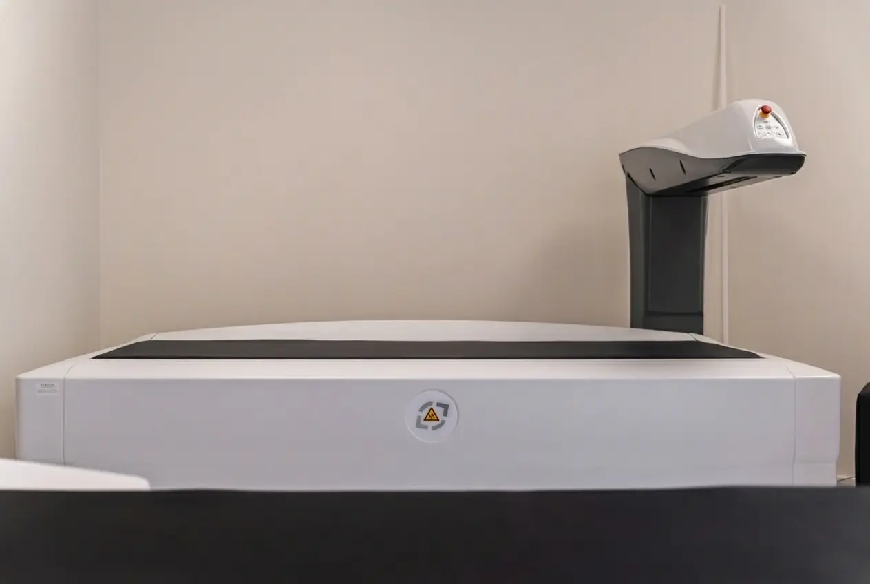
The exam is done without an injection, under an open machine. You keep still during each pass of the scanning arm, then leave straight away. In total, allow about 15 minutes.
After the exam
After the exam: report and images
A radiologist interprets the measurements and writes a report for your doctor. The report is also published automatically on the Brussels Health Network and on MyHealth (Ma Santé). They explain what the figures mean for your situation; your doctor decides the next step with you. The result is available within 24 hours. The report gives your bone scores and, if an earlier measurement exists, how they have changed.
Your doctor places these figures in your history (age, fractures, treatments) and decides with you on a treatment or a check-up.
As soon as the radiologist has interpreted the exam, your images and the report appear on the PACSonWEB portal, with the access code the secretary gives you on site.
If a fracture or a collapsed vertebra is suspected, the doctor sometimes requests another exam: see the related exams.
Your questions
Frequently asked questions about bone density scans
Is a bone density scan reimbursed?
Yes, under conditions set by the INAMI (RIZIV), which depend on your medical situation and on the time since your last measurement. Your doctor checks that you meet them; your health insurance fund tells you what you pay.
What do the T-score and Z-score mean?
The T-score compares your bone density with that of a young adult: at -1 and above it is normal, between -1 and -2.5 the bone is slightly weakened (osteopenia), at -2.5 and below, osteoporosis. The Z-score compares it with people of your age.
How often should the scan be repeated?
Your doctor sets the pace according to the result and the treatment. A check after about two years is common, sooner with some treatments such as high-dose corticosteroids.
Should I come back to the same center?
It is better: two measurements compare best on the same machine. The change in your figures from one scan to the next is then more reliable than a single figure.
Read on
Exams related to the bone density scan
Depending on the result or the question asked, the doctor sometimes adds another exam to the bone density scan.
- DEXA body composition scanMeasures fat, muscle and bone region by region, without a prescription.
- X-rayShows a fracture or collapsed vertebra that the bone density scan only measures in figures.
- MammographyThe X-ray of the breast, done at all our centers.
- MRI and CT scanOrganized by Access Imaging at our partner hospitals; our team arranges the appointment for you.
Find all our exams on the exams page, addresses on the centers page, more answers in the frequently asked questions, or write to us through the contact page.